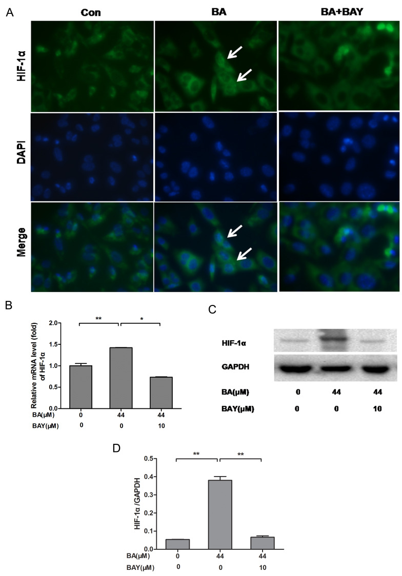
Figure 3.
BA promotes the expression of HIF-1α in newborn-mouse cartilage chondrocytes and this effect was reversed by BAY. (A) HIF-1α nuclear localization in BA (44 µM)-treated chondrocytes with or without BAY (10 µM) was observed under a fluorescence microscope (magnification, 400x), the white arrows indicate chondrocytes with strong nuclear HIF-1α expression. (B-D) mRNA and protein levels of HIF-1α measured by RT-qPCR and western blotting, respectively. β-actin was the control for RT-qPCR and GAPDH was the control for western blotting. *P<0.05, **P<0.01. HIF-1α, hypoxia-inducible factor-1α; BA, baicalin; BAY, hypoxia inducible factor-1α inhibitor (BAY-87-2243); RT-qPCR, reverse-transcription quantitative; con, control; BAY, hypoxia inducible factor-1α inhibitor.

