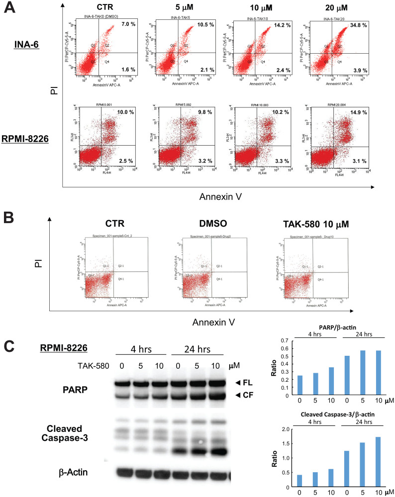
Figure 2. TAK-580 induces apoptosis in MM cells.
(A) INA-6 cells were treated with TAK-580 (0–20 μM) for 24 h; RPMI-8226 cells were treated with TAK-580 (0–20 μM) for 48 h. Apoptotic cells were analyzed with flow cytometry using annexin V/PI staining. Apoptosis was assessed as the percentage of annexin V-positive cells. (B) Normal donor peripheral blood B lymphocytes were treated with dimethylsulfoxide or TAK-580 (10 μM) for 48 h. Apoptotic cells were analyzed with flow cytometry using annexin V/PI staining. Apoptosis was assessed as the percentage of annexin V-positive cells. (C) RPMI-8226 cells were treated with TAK-580 (0–10 μM) for 4 or 24 h. Whole-cell lysates were subjected to western blotting using PARP, cleaved caspase-3, and β-Actin Abs. FL, full-length; CF, cleaved form. (Upper right panel): The graph represents ratios of PARP CF density relative to β-Actin in Figure 2C. (Lower right panel): The graph represents ratios of cleaved caspase-3 density relative to β-Actin in Figure 2C.

