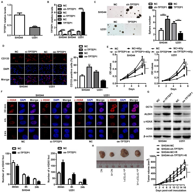
Figure 2.
TPTEP1 suppresses glioma cell stemness and radioresistance in vitro and in vivo. RT-qPCR analysis was performed to detect TPTEP1 expression levels in (A) SHG44 and U251 cells and in (B) stable/transient TPTEP1-depleted SHG44, stable TPTEP1-overexpressed U251 cells and the control cells. (C) Tumorspheres formation assay was performed in stable TPTEP1-depleted SHG44 cells, stable TPTEP1-overexpressed U251 cells and the control cells (×100 magnification). (D) Immunofluorescence analysis was performed to detect the expression and location of CD133 in transient TPTEP1-depleted SHG44 cells, transient TPTEP1-overexpressed U251 cells and their corresponding controls (×400 magnification). (E) Cell proliferation and radiosensitivity assays were performed in transient TPTEP1-depleted SHG44 cells, transient TPTEP1-overexpressed U251 cells and their corresponding controls. (F) Immunofluorescence analysis was performed to evaluate γ-H2AX foci formation in transient TPTEP1-silenced SHG44 cells, transient TPTEP1-overexpressed U251 cells and their corresponding controls. (G) Western blot analysis was performed to detect OCT4, ALDH1, γ-H2AX and H2AX expression levels in transient TPTEP1-silenced SHG44 cells, transient TPTEP1-overexpressed U251 cells and their corresponding controls. (H) A subcutaneous transplantation tumor model was applied to assess glioma cell proliferation regulated by TPTEP1 and R. *P<0.05, **P<0.01, ***P<0.001. TPTEP1, transmembrane phosphatase with tensin homology pseudogene 1; RT-qPCR, reverse transcription-quantitative PCR; OCT4, Octamer binding protein 4; ALDH1, aldehyde dehydrogenase 1; γ-H2AX, Histone H2AX; R, radiation.

