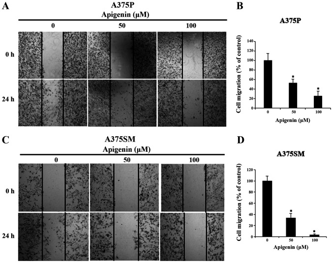
Figure 3.
Effect of apigenin on melanoma cell migration ability. (A) A375P and (C) A375SM melanoma cells were treated with apigenin (0, 50, 100 µM) for 24 h and cell density in the wound area was measured using a wound healing assay (magnification, ×100). (B and D) The percentage of cell migration was estimated compared with the cell density in the wound area of the untreated control group. Cell density in the wound area for any field of view was measured at different locations. Data are presented as the mean ± standard deviation from three independent experiments. Significance was determined using ANOVA followed by a Dunnett's post hoc test. *P<0.05 vs. untreated control group.

