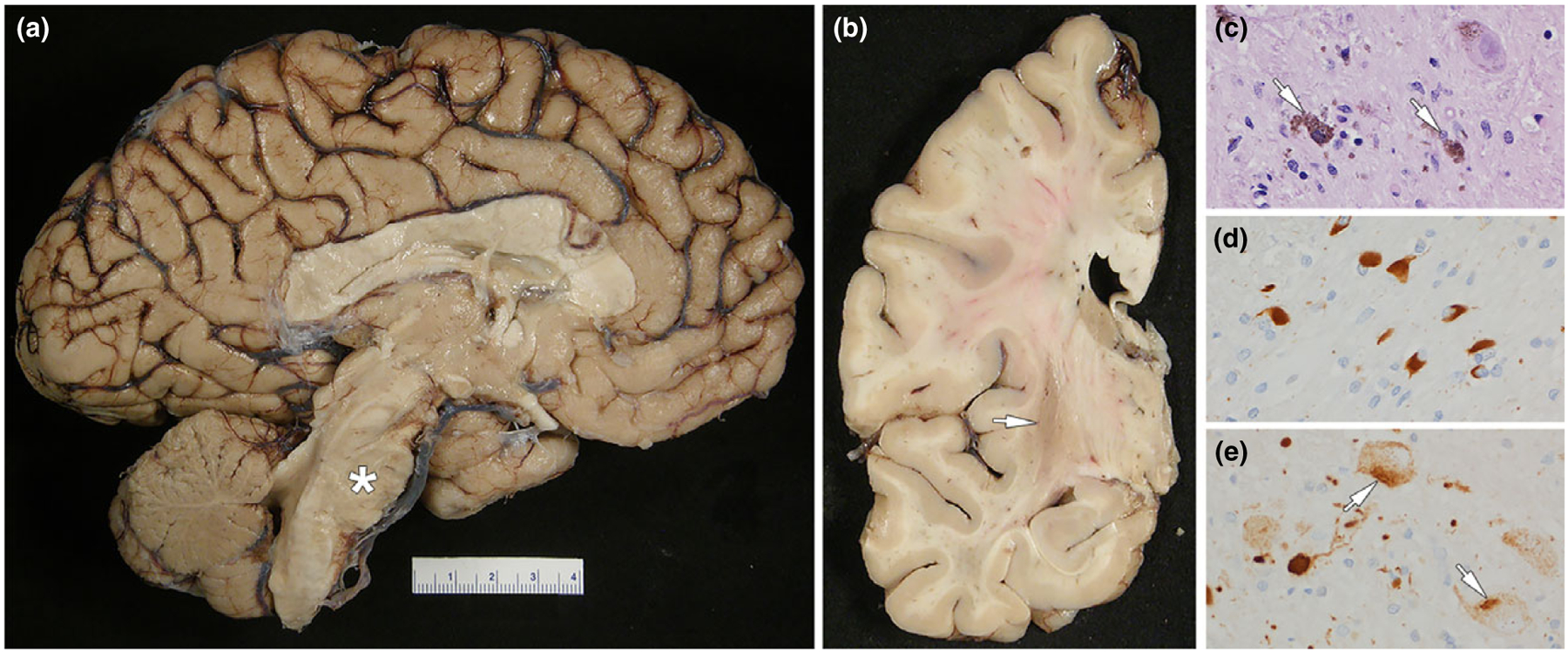
Figure 2.

MSA: (a) medial view of the left cerebral hemisphere showing severe atrophy of the pontine base (asterisk); note also the cerebellar vermis atrophy and discoloration; (b) coronal section of the left cerebral hemisphere showing atrophy and dark brown discoloration of the posterior putamen (arrow); (c) neuronal loss in substantia nigra with neuromelanin containing macrophages (arrows) (d) fiber tracts in the pontine base have numerous oligodendroglial inclusions (GCIs) with α-synuclein immunohistochemistry; (e) neurons in the pontine base have neuronal cytoplasmic inclusions (arrows) with α-synuclein immunohistochemistry (note also the dystrophic neurites). The patient had MSA with a mixed subtype since age 55 years. She had limb ataxia, rigidity, urinary incontinence and anxiety. Further, rapid eye movement sleep behavior disorder and cognitive decline developed. Age at death was 64 years.
