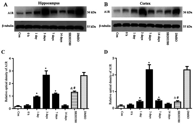
Figure 2.
A1R protein was detected by western blot analysis. The relative optical density of A1R protein band was normalized to that of β-tubulin. A1R protein expression levels in the (A) hippocampus and (B) cortex of epileptic and con rats. Bar graphs for (C) hippocampus and (D) cortex of epileptic and con rats. Compared with con rats, A1R protein expression levels in the hippocampus of rats at 1 and 3 days and 7 days after seizure induction were upregulated. Compared with the con group, A1R protein expression levels were not significantly different at 0 h and 14 days. SB203580 inhibited A1R protein expression levels compared with epileptic rats at 3 days after seizure induction, whereas DMSO did not have this effect. n=5 in each group. *P<0.05 vs. con; #P<0.05 vs. DMSO; &P<0.05 vs. 3 days. A1R, adenosine A1 receptor; con, control.

