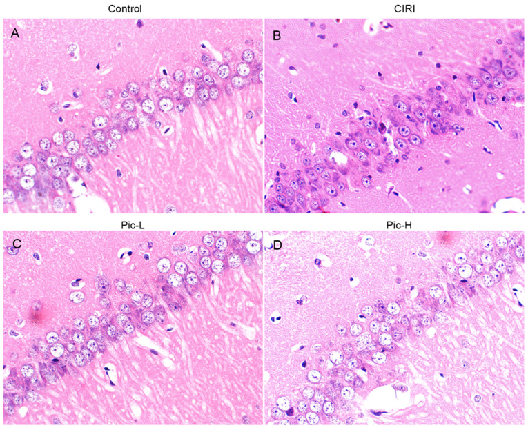
Figure 2.
Histological analysis of the effects of Pic on neuronal injury induced by CIRI in mice. Hematoxylin and eosin staining was performed on tissue sections from the hippocampal Cornu Ammonis 1 regions (magnification, ×400; n=5 per group). (A) Neurons with normal histology were observed in the Sham group. (B) Altered neurons characterized by nuclear pyknosis, neurons decrease and neuron atrophy were found in the CIRI group. (C) Neuronal alterations were slightly eliminated in the Pic-L treatment group. (D) Neuronal alterations were significantly eliminated in the Pic-H treatment group. CIRI, Cerebral ischemia-reperfusion injury; Pic, Piceatannol; L, low; H, high.

