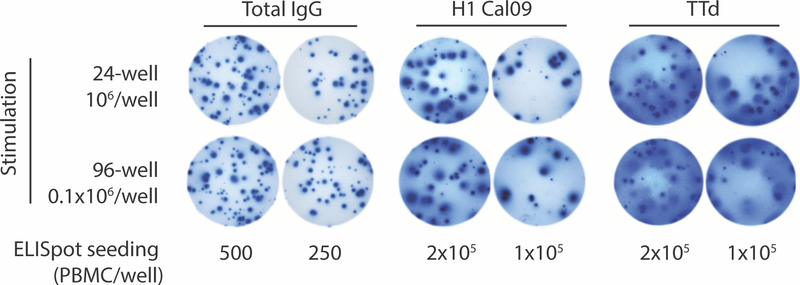
Figure 3.
A portion of a developed ELISpot plate used to enumerate total and antigen-specific IgG MASCs after in vitro MBC stimulation. PBMCs from a healthy donor were stimulated and analyzed by ELISpot assay as described in Basic Protocols 1 and 2. Wells in the ELISpot plate were coated with goat anti-human IgG for measurement of total IgG MASCs and with influenza virus HA (H1 Cal09) or tetanus toxoid (TTd) for measurement of MASCs specific for those antigens. Note the differences in spot size and appearance. This can reflect the amount of IgG secreted by individual MASCs and the affinity of the interaction between secreted IgG and the anti-IgG capture Ab or the target antigens. The quality of well coating is also an important determinant of spot appearance. True spots are typically round with a darker center and a faint edge, reflecting less Ab with distance from the MASC.

