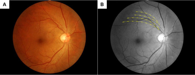
Fig 1. RNFLDs with their central ends around the retinal vessels.
Representative fundus photos of non-glaucomatous RNFLDs with their central ends around the retinal vessels. Yellow dotted lines indicate the RNFLDs. This case is from the PA group and shows two RNFLDs with their central ends adjacent to the superior retinal vein and artery. (A) Color photo, (B) Red-free image.

