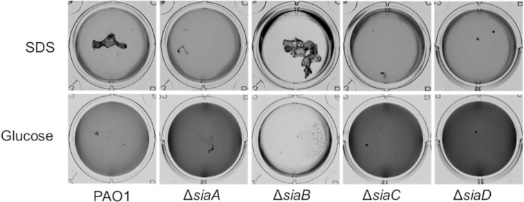
Fig 1. Growth of the P. aeruginosa strains on 3.5 mM SDS or 22 mM glucose in 12 well microtiter plates after 18 h, at 30°C and 200 rpm.
Images were acquired using a flat-bed scanner (Umax Powerlook), normalized by the “match colour” function of Photoshop (Adobe Photoshop CS5), and converted to greyscale.

