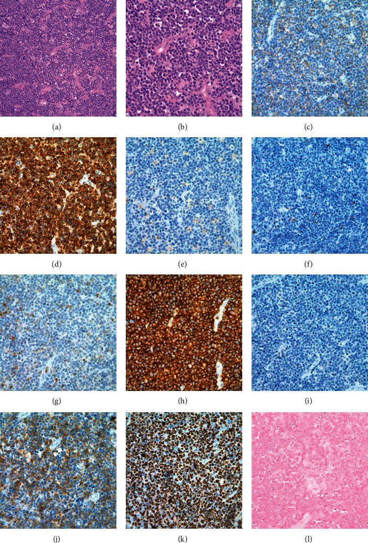
Figure 2.

Biopsy of the left cervical lymph node. Hematoxylin and eosin (a, b), immunohistochemistry staining (c–j), and in situ hybridization (l). (a, b) H&E sections showing sheets of pleomorphic atypical medium-to-large lymphocytes. (c) CD2. (d) CD3. (e) CD4. (f) CD5. (g) CD7. (h) CD8. (i) CD20. (j) CD25. (k) Ki67. (l) Epstein–Barr virus- (EBV-) encoded RNAs (EBER) (original magnification, ×200 (a, c–l) and ×400 (b)).
