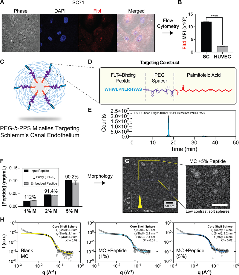
Figure 1.
The design of PEG-b-PPS micelles that target SC cells in the anterior segment of the eye. A, B) Human SC cells express Flt4/VEGFR3. A) Representative confocal microscopy images of SC endothelial cells that were stained with anti-FLT4 antibody to confirm presence of FLT4 on the cell surface. Cells were stained with Hoescht 33342 (blue) and FLT4 (red) B) Flow cytometry data comparing median fluorescence intensity (MFI) of SC cells and HUVECs stained with anti-FLT4 antibodies. Data shown as mean±SEM. Significance determined by unpaired t-test (****p<0.0001). C) Schematic representation of peptide-displaying micelles. D) The peptide targeting construct consisting of targeting peptide, PEG spacer and palmitoleic acid tail. E) LCMS spectra of the purified Flt4-targeting peptide construct. F) Peptide loading efficiency of PEG 6 peptide construct into PEG-b-PPS micelles at various molar ratios as determined by tryptophan fluorescence measurements. Data shown as mean ±SD, N=3 technical replicates. G) STEM micrograph of negatively stained PEG-b-PPS micelles (MC) displaying the peptide targeting construct (5%) (200,000X magnification). H) Synchrotron small angle x-ray scattering (SAXS) plots for Blank micelles, and micelles displaying the targeting peptide at 1% and 5% molar ratios. A core shell model (solid line) was fit to the data (gray points). The core radius (rc), shell thickness (rt), and total MC radius (r) is displayed together with the chi square (X2) value for the final model fit.

