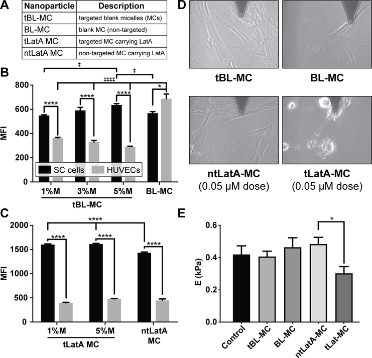
Figure 2.
The micellar display of a FLT4-binding peptide targets the delivery of Lat A to SC endothelial cells in vitro. A-C) The FLT4-targeting peptide enhances micelle uptake by SC endothelial cells and decreases uptake by HUVEC. A) Nanoparticle formulation abbreviations. B) unloaded micelles, C) micelles loaded with latrunculin (LatA). Cells were incubated for 1 h with Alexa 555 labeled micelles incorporated with various molar ratios (1%, 3%, 5%) of peptide to micelle. After 1 h incubation, cells were washed, harvested and fixed for analysis via flow cytometry. Median fluorescence intensity (MFI) was measured to quantify uptake of the various formulations by both cell types. Data shown as mean ± SEM (n=3 biological replicates). In B, significance determined by one way ANOVA for each of the two cell types with post hoc Tukey’s multiple comparisons test (‡‡‡‡ p<0.0001, ‡p<0.05). In B, C)Two way ANOVA was used to evaluate differences between all groups with post hoc Sidak’s multiple comparisons test (****p<0.0001, *p<0.05). D-E) Targeted delivery of Lat A decreases the stiffness of human SC cells. D) Representative images taken during AFM measurements of cells treated for 2 hours with BL-MC, tBL-MC (0.97 mg/mL), ntLatA-MC, or tLatA-MC (0.05 μM Lat A, 0.97 mg/mL); AFM tip is at top of each panel. E) Stiffness of SC cells after 2 h was determined by AFM; data shown is geometric mean ± SD (5 measurements/condition). In E), significance determined by ANOVA with post hoc Tukey’s multiple comparisons test (*p<0.05).

