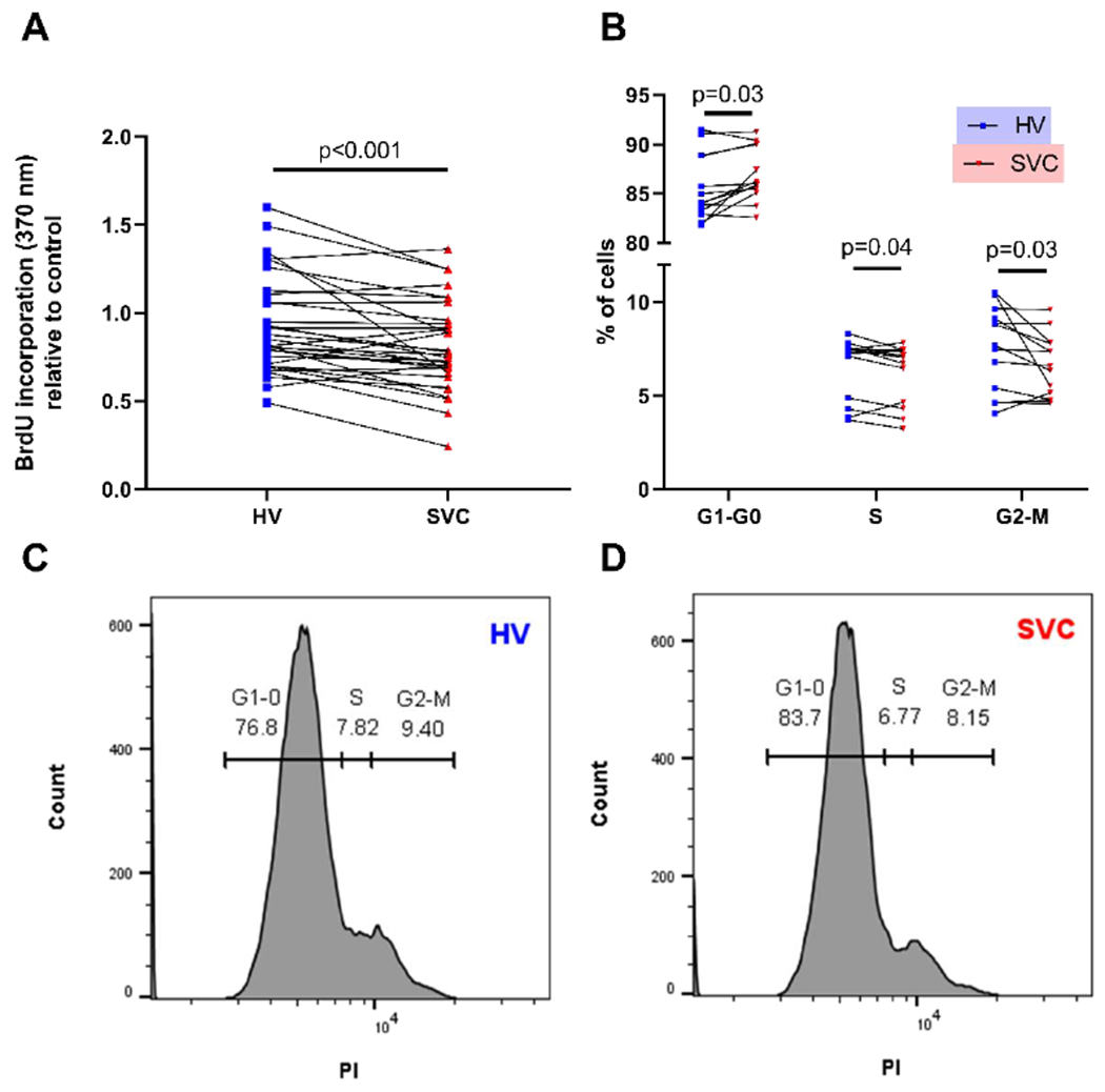
Figure 2:

HV serum increases HPMEC proliferation compared to SVC serum. (A) Quantification of HPMEC proliferation using colorimetric absorbance of BrdU incorporation. N=32 per group. Before and after plot represents individual patient samples with lines connecting paired patient samples. (B) Quantification of HPMEC proliferation using cell cycle analysis with PI as detected by flow cytometry. N=13 per group. Before and after plots represent individual patient samples with lines connecting paired patient samples. (C-D) Representative histograms of cell cycle analysis. HPMEC= human pulmonary microvascular endothelial cells, HV= hepatic vein, PI=propidium iodine, SVC= superior vena cava.
