
FIGURE 7.
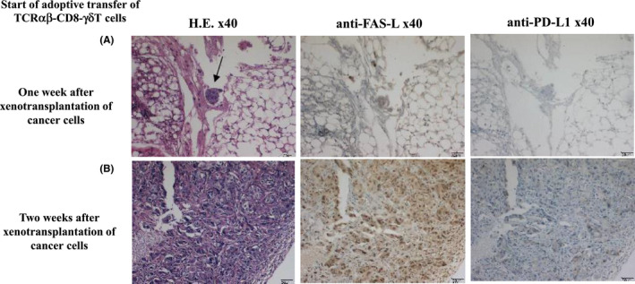
There was no effect of adoptively transferring TCRαβ‐CD8 γδT cells starting 2 wk after xenotransplantation of cancer cells (B901L‐HLA‐B15), so we analyzed the xenotransplanted cancer cells. Immunohistochemical staining of the xenotransplanted cancer cells using CD3, PD‐L1, FasL, VEGF, transforming growth factor‐β (TGF‐β) and HLA class I was performed. Intravenous injection of TCRαβ‐CD8 γδT cells was started 1 wk after xenotransplantation of B901L‐HLA‐B15 in (A) and 2 wk after xenotransplantation in (B). The difference between the 2 groups was seen in expression of FasL but not PD‐L1 expression