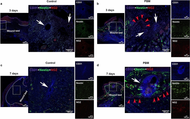
Figure 7.
PBM effects on the skin microvasculature. CD31 marker epifluorescence images of the immunostaining for control and PBM-treated wounds at day 3 (a,b) and 7 post-surgery (c,d). In all groups and experimental times, the squared region is depicted in higher magnification (× 200) in the following image. PECAM-1 positive vessels (white arrows) are observed in the control (a,c) and in the PBM-treated samples (b,d) in both experimental times. Note the presence of an arteriole in a representative PBM sample (d). Also, note small capillaries in the photoactivated samples where the NG2 protein is observed colocalized with the CD31 marker (red arrowheads) (b,d). Scale bar = 100 µm.

