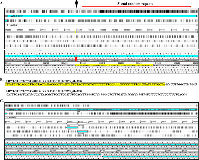
FIG 4.
Himar1 Tn insertion site in the A. phagocytophilum virB6-4 gene. (A) Artemis (genome browser and annotation tool; Wellcome Sanger Institute, UK) window showing A. phagocytophilum wild-type virB6-4 gene used as a reference for the location of the Himar1 insertion site. The first panel shows a “zoomed out” view of the virB6-4 DNA sequence translated into six reading frames with black bars indicating stop codons. The open reading frame (ORF) for virB6-4 is shown in light blue. The black arrow indicates the Himar1 insertion location, 5′ upstream the 3′ end tandem repeats. The yellow box is “zoomed in” in the panel below to show in detail the virB6-4 sequences flanking the Himar1 Tn (yellow highlight), including the TA dinucleotide site required for successful transposition (red arrow). (B) Paired-end DNA sequencing reads indicating the same A. phagocytophilum sequences as described above (yellow highlight) and Himar1 Tn sequences (italics). (C) Artemis window of in silico cloning of the Himar1 Tn in the opposite orientation to virB6-4. Black bars within the virB6-4 ORF indicate putative stop codons.

