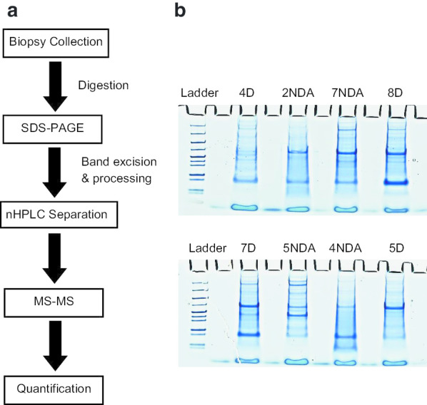
Fig. 1.

Experimental workflow of IC bladder biopsies. a Patient biopsies were digested and separated by SDS-PAGE before analysis via nHPLC-MS/MS. Data was searched using Mascot, and a list of detected proteins was created for each sample. Significant proteins were mapped for associations and shared function via STRING. b 10 μg of digested bladder tissue was loaded onto an SDS-PAGE and separated. Bands were excised and digested prior to nHPLC-MS/MS. Samples were blinded and randomized to avoid bias. Patient number corresponds to patient number in Table 1. D = Disease tissue biopsy; NDA = Non-disease apparent tissue biopsy
