Fig. 4.

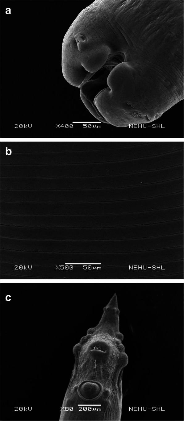
Scanning electron micrographs of an untreated A. galli. a The anterior part bears three terminal blob-like structures called the lips, which have small protrusion called papilla (upper arrow) and surround the mouth (lower arrow). × 400. Scale bar = 50 μm. b The body surface called the cuticle in the middle of the body is smooth and lined with parallel rows of transverse striations called annulations (arrows). × 500. Scale bar = 50 μm. c The posterior end is the tail which bears anal opening or cloaca (top arrow), a precloacal sucker (middle arrow) and sensory protrusions called phasmids (bottom arrow). × 80. Scale bar = 200 μm
