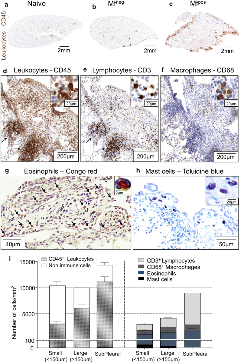
Fig. 5.
Polyps show a strong immune infiltration. Lungs were recovered from naive, Mfneg and Mfpos gerbils at 70 days p.i. Immune cells were stained in 4 µm thick sections. a–f Immunostainings were performed for leukocytes (CD45), lymphocytes (CD3) and macrophages (CD68). Overview of lungs section from naive (a), Mfneg (b) and Mfpos (c) gerbils stained with CD45 (brown). d–f View of a large polyp stained for CD45 (d), CD3 (e) and CD68 (f). Arrows show subpleural inflammatory foci. g Eosinophils were stained with Congo red; arrows indicate eosinophils. h Mast cells were stained with Toluidine blue. In all images, the right quadrant shows a zoom of positive cells. i Quantification of cell concentrations in small polyps (< 150 µm), large polyps (> 150 µm) and subpleural infiltrates. Results are expressed as the mean ± SEM (n = 7 Mfpos gerbils for CD45, CD3, CD68 and toluidine blue staining and 4 for Congo Red)

