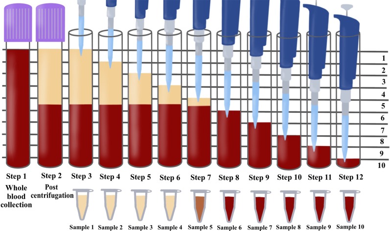
Fig. 2.
Illustration demonstrating the proposed novel method to quantify cell types following the centrifugation of PRF. One of the limitations of the current methods utilized to investigate PRF cellular content from the whole plasma layer is the inability to accurately determine where cells migrate following centrifugation. By utilizing the proposed technique using the sequentially pipetting 1 mL layers from the top layer downwards, it is possible to quantify cell numbers in 10 samples from CBC analysis and accurately determine the precise location of each cell layer following centrifugation at various protocols. Note that one layer (in this case, layer 5) will contain both yellow plasma and red blood cells. This effect is figuratively depicted with arrows to demonstrate the location of the buffy coat where a higher concentration of platelets/leukocytes is typically found.
Reprinted with permission [16]

