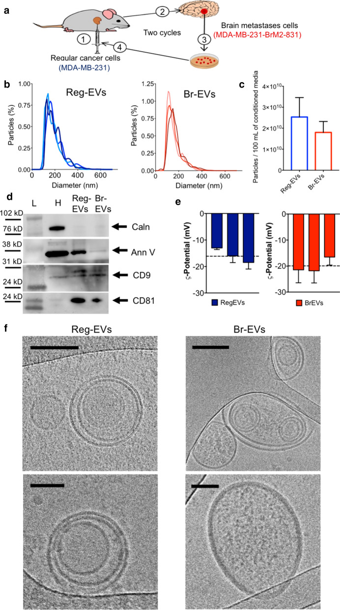
Fig. 1.
Characterization of extracellular vesicles (EVs) derived from regular (Reg) and brain metastases (Br) breast cancer cells. The conditioned media of regular human MDA-MB-231 breast cancer cells and a brain metastases variant of MDA-MB-231 cells (MDA-MB-231-BrM2-831) were processed by tangential flow filtration to obtain Reg-EVs and Br-EVs, respectively. a Schematic of in vivo selection cycles to obtain a brain metastases cell line: (1) intracardiac injection of cells, (2) development of brain metastases, (3) excision of brain metastases and in vitro culture, and (4) intracardiac injection of cultured cells. b Size distribution profiles (10 nmincrements) measured by nanoparticle tracking analysis (NTA). Data are shown as biological triplicates. c Particles isolated from 100 mL of conditioned cell culture media. d Protein markers of EVs (annexin V, cluster of differentiation (CD)9, and CD81) and intracellular contaminants (calnexin) obtained by Western blot. Arrows represent the expected protein band. e Zeta potential measured by laser Doppler micro-electrophoresis. Data are presented as mean ± SD of three technical replicates. Dashed line represents the mean value of three biological replicates. f Representative cryogenic transmission electron microscopy images. Scale bar, 100 nm (upper panels) and 50 nm (lower panels). Ann V annexin V, Caln calnexin, H cell homogenate; L protein ladder

