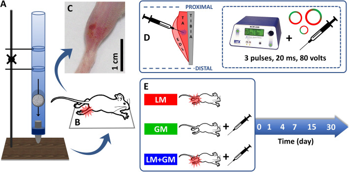
Fig. 1.
Illustration of skeletal muscle injury and gene therapy. (a) The height of the device was adjusted to 100 cm to transfer the 16.2-g projectile to the (b) tibialis anterior (TA) muscle of the animal placed in the supine position. (c) Representative image of an injured leg after 24 h and (d) representative scheme of the injection of the plasmid vectors into the central portion of the TA muscle; the overlapping structures, extensor digitorum longus (EDL), and tibial bone are also shown. (e) Experimental timeline and animal groups: GM, healthy mouse treated with GM-CSF; LM, injured mouse without treatment; LM+GM, injured mouse treated with GM-CSF

