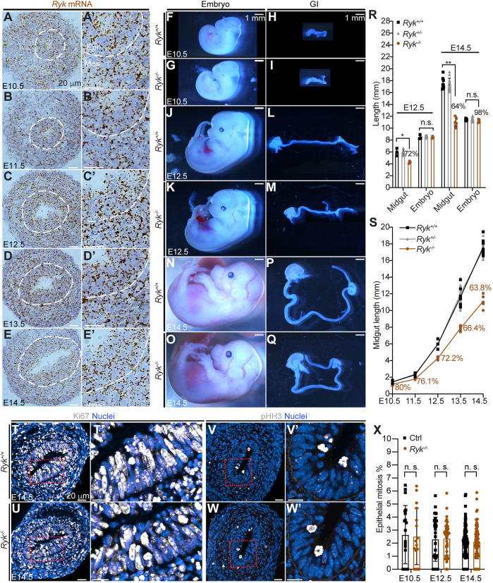
Fig. 3.
RYK is required for midgut elongation throughout Phase I. (A-E′) RNAscope of Ryk on sections of wild-type midguts at E10.5-E14.5. (A′-E′) Higher magnifications of A-E. The epithelial-mesenchymal interface is outlined by white dashed lines. Epi, epithelium; Mes, mesenchyme. Scale bars: 20 µm. (F-Q) Embryos and GI tracts of the wild-type (F,H,J,L,N,P) and Ryk null (G,I,K,M,O,Q) at E10.5, E12.5 and E14.5. Scale bars: 1 mm. (R) Quantitation of midgut length and embryo length of Ryk+/+ (black), Ryk+/− (gray) and Ryk−/− (brown) at E12.5 and E14.5. At E12.5, Ryk+/+, n=3; Ryk+/−, n=5; Ryk−/−, n=3. At E14.5, Ryk+/+, n=8; Ryk+/−, n=14, Ryk−/−, n=5. (S) Dynamics of midgut lengthening in Ryk+/+ (black), Ryk+/− (gray) and Ryk−/− (brown) embryos from E10.5 to E14.5. Ryk+/+: n=5 (E10.5), n=4 (E11.5), n=3 (E12.5), n=7 (E13.5), n=8 (E14.5); Ryk+/−: n=7 (E10.5), n=6 (E11.5), n=5 (E12.5), n=12 (E13.5), n=15 (E14.5); Ryk−/−: n=3 (E10.5), n=3 (E11.5), n=3 (E12.5), n=5 (E13.5), n=5 (E14.5). (T-W′) Immunostaining for Ki67 (T-U′) and pHH3 (V-W′) on cross-sections of the central Ryk+/+ and Ryk−/− midgut at E14.5. (T′,U′,V′,W′) Higher magnification of boxed areas in T,U,V,W, respectively. Scale bars: 20 µm. (X) Quantitation of mitosis rate (% pHH3 cells) in the epithelium of control and Ryk−/− midguts. Measurements were performed on 20 sections of five control samples and 16 sections of four Ryk−/− samples at E10.5; 30 sections of three control samples and three Ryk−/− samples at E12.5; 40 sections of five control samples and five Ryk−/− samples at E14.5. Data are mean±s.e.m. Analyses were performed using unpaired nonparametric tests (Mann–Whitney test). *P<0.05, **P<0.01; n.s., not significant.

