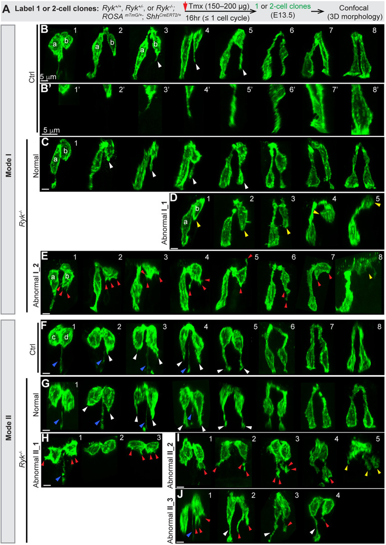
Fig. 5.
In the absence of RYK, some ‘pathfinding’ daughter cells fail to grow a basally directed filopodial protrusion to make a basal connection. (A) Strategy for generating one- or two-cell clones in control and Ryk−/− midgut epithelial tubes at E13.5 for confocal imaging. (B-E) 3D reconstructions of Mode I daughter pairs in control (B,B′) and Ryk−/− (C-E) midguts. Normal basally oriented filopodial protrusions (B,C) are indicated by white arrowheads. B′ shows higher magnifications of the bottom regions of pathfinding cells shown in B: the basal tip of recently divided cells (1′,2′); early filopodial projection (3′,4′); filopodial thickening and basal connection (5′-8′). In D, cells with no filopodial protrusion are indicated by yellow arrowheads. In E, abnormal protrusions are indicated by red arrowheads; the yellow arrowhead in E8 indicates a pre-apoptotic cell. Scale bars: 5 µm. (F-J) 3D reconstructions of Mode II pairs in control (F) and Ryk−/− (G-J) midguts. Normal basally oriented filopodial protrusions are indicated by white arrowheads in F,G,J; abnormal protrusions are indicated by red arrowheads in H-J; the inherited basal processes are indicated by blue arrowheads in F-H,J. The yellow arrowheads in I5 indicate a fragmented cell. Scale bars: 5 µm.

