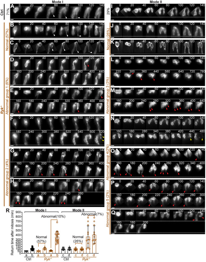
Fig. 6.
RYK facilitates efficient basal tethering of ‘pathfinding’ cells, which is essential for a timely nuclear return. (A-Q) Live imaging of the basal return of Mode I (A-H) and Mode II pairs (I-Q) in control (A,I) and Ryk−/− midguts (B-H; J-Q). Sequential 2D images taken at labeled time points (minutes) are displayed; time 0 marks the mitosis. White arrowheads indicate normal filopodial protrusions; red arrowheads indicate abnormal protrusions; yellow arrowheads indicate apoptotic cells. White dotted lines mark the basal surface. Scale bars: 10 µm. (R) Quantitation of nuclear return time for Mode I daughter a and b, and Mode II daughter c and d in control (black) and Ryk−/− (brown) midguts. Solid squares and circles reflect the actual returning time. Open squares and circles reflect the time that nuclei stay at the apical side during the recording. Ninety-one daughter pairs from five control midguts and 176 pairs from six Ryk−/− midguts were analyzed. Data are mean±s.e.m.

