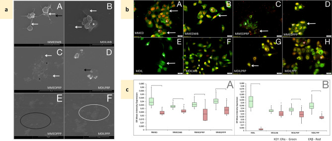
Figure 3.
Effects of blood constituent incubation with MCF7 cells—controls. MMED media-treated MCF7 cells, MMEDWB WB co-incubated with media-treated MCF-7 cells, MMEDPRP PRP co-incubated with media-treated MCF-7 cells, MMEDPPP PPP co-incubated with media-treated MCF-7 cells, MDIL diluent-treated MCF7 cells, MDILWB WB co-incubated with diluent-treated MCF-7 cells, MDILPRP PRP co-incubated with diluent-treated MCF-7 cells, MDILPPP PPP co-incubated with diluent-treated MCF-7 cells. (a) Platelet ultrastructural alterations. White*—membrane folds, black*—hyalomere spread, white arrow—extending filipodia, black arrow—microparticles, white circle—fibrin, black circle—fibrin pores A: MMEDWB—active platelets, smooth membrane, extending filipodia and microparticles. B: MDILWB—platelets with membrane folds and filipodia. C: MMEDPRP—spread platelets and filopodia. D: MDILPRP—platelets with membrane folds and microparticles. E: MMEDPPP and F: MDILPPP—remnants of platelets, fibrin and pores. (b) Co-localisation of ERα (green) and ERβ (red) in MCF7 cells, following exposure to blood constituents. A: MMED—primarily cytoplasmic (white arrow) ERα and nuclear (*) ERβ expression. B: MMEDWB—cellular processes extend, cytoplasmic ERα and nuclear ERβ. C: MMEDPRP—diffuse ERα and ERβ expression. D: MMEDPPP—high cytoplasmic ERα expression and ERβ contained within the nucleus. E: MDIL—nuclear and cytoplasmic ERα, some nuclear ERβ. F: MDILWB—nuclear and cytoplasmic ERα, greater nuclear ERβ expression. G: MDILPRP and H: MDILPPP—high ERα and ERβ nuclear expression, minimal cytoplasmic expression. (c) Box and whisker plots representing quantitative analysis of ERα (green) and ERβ (red) expression in MCF7 cells, following exposure to blood constituents. A: Conditioned media-treated MCF7 cells. B: Diluent-treated MCF7 cells (0.1% DMSO). [p < 0.05 between ERα and ERβ within the treatment group. *p < 0.05 between matched ERs compared to untreated MCF7.

