Figure 3.

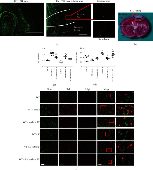
Optimized integration of TP and Ki20227 pretreatments correspondingly increases and decreases NeuN and Iba1 expressions in the cortical penumbra. (a) Thy-YFP mice were employed to show the ischemia core and penumbra region. We counted fifty YFP-positive cells and axons in the pyramidal neurons. After stroke, YFP-positive cell deaths with fluorescence were suppressed in similitude to the normal core region. Scale bar = 100 μm and 50 μm. (b) TTC staining 24 hours after ischemia showing the ischemic core and peri-infarct region. The pink area between the normal cortex and ischemic core was the penumbra region. (c) Quantitative analyses of Iba1 expression. (d) Quantitative analyses of NeuN expression. (e) Immunofluorescence staining images of experimental groups. Scale bar = 20 μm. Data were expressed as mean ± SEM. One-way ANOVA with Tukey's tests, n = 5. The WT vs. WT+stroke groups, ∗∗∗P < 0.0001; the WT+K vs. WT+stroke+K groups, ∗∗∗P < 0.0001; the WT+stroke group vs. WT+stroke+TP and WT+stroke+K groups, #P < 0.05, ##P < 0.01; the WT+K group vs. WT group, &P < 0.05; and the WT+stroke+K+TP group vs. WT+stroke+TP and WT+stroke+K groups, ^P < 0.05, ^^P < 0.01.
