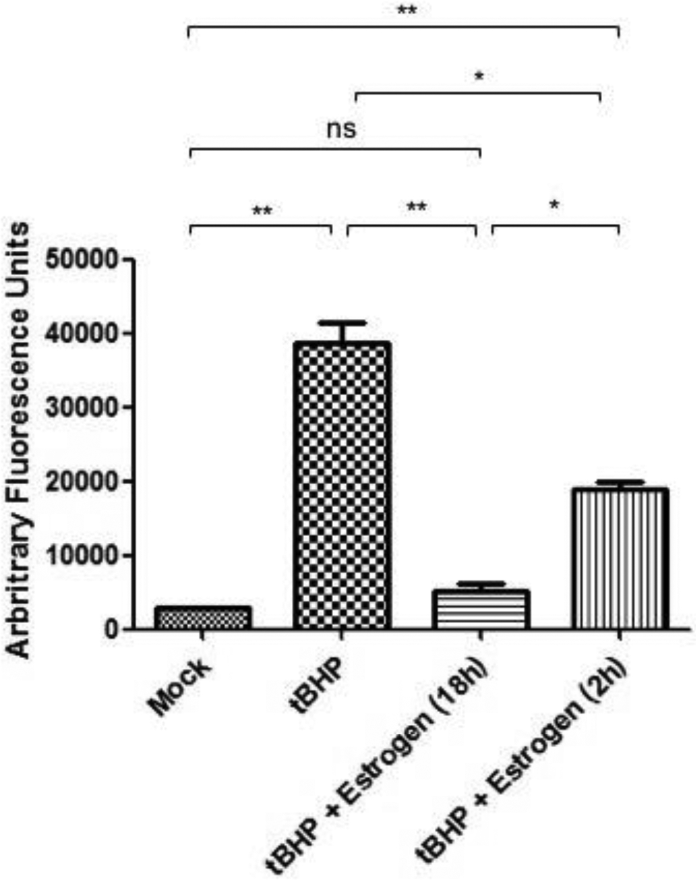
Figure 2. Estrogen inhibits oxidative stress induced caspase activation in ONHAs.

Oxidative stress in ONHAs were induced using tBHP and caspase activity was measured using Ac-DEVD-AFC. Caspase activity in ONHAs that were treated with tBHP was significantly higher compared to mock treated (**, p=0.0058). ONHAs pretreated with 25 μM estrogen for 2 h (*, p=0.0208) or 18 h (**, p=0.0077) before tBHP addition had a significant decrease in caspase activity compared to tBHP treated cells. Estrogen pretreatment for 18 h was significantly more effective at inhibiting caspase activity compared to a 2 h pretreatment (*, p=0.0144). ONHAs pretreated for 2 h with estrogen had a significant level of active caspases versus mock treated (**, p=0.0035) while pretreatment with estrogen for 18 h showed no significant difference in caspase activity versus mock treated (ns, p=0.1974). Experiments were done in triplicate (n=3) and values were depicted as mean +/− SEM and analyzed using ANOVA (p=0.0003). For statistical comparison, the Bonferroni post-hoc test and Student t-test were used. Statistical significance was set at p ≤ 0.05.
