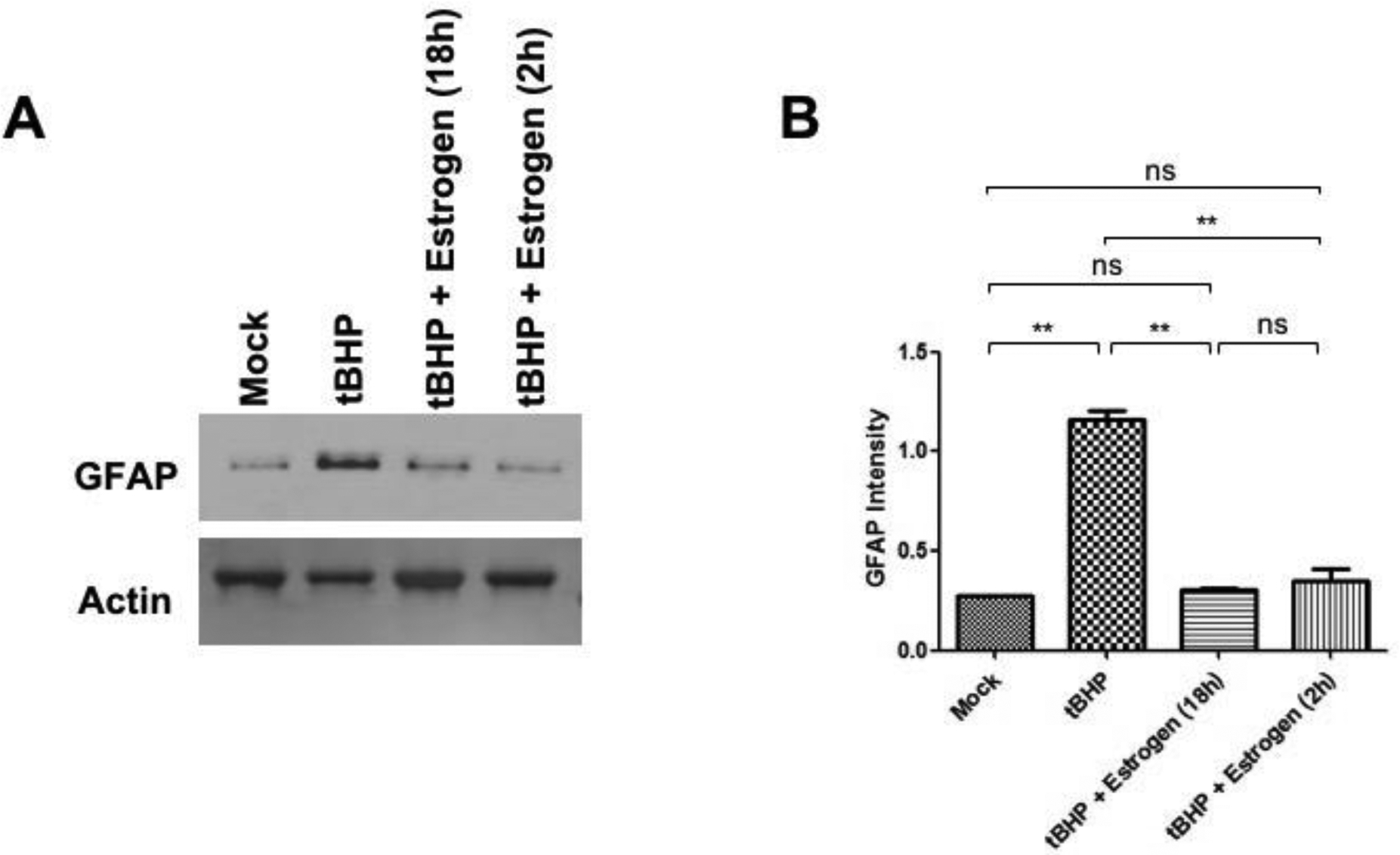
Figure 6. During oxidative stress induced by tBHP estrogen stabilizes GFAP levels in optic nerve head astrocytes (ONHAs).

(A) GFAP levels are elevated during oxidative stress as determined by immunoblot assay (B) During oxidative stress GFAP levels are significantly upregulated versus mock (**, p=0.0020). 25 μM estrogen pretreatment for 18 h (**, p=0.0022) or 2 h (**, p= 0.0074) significantly reduced GFAP levels compared to tBHP treated ONHAs. When compared to mock treated ONHAs both 2 h (ns, p=0.3054) and 18 h (ns, p=0.0954) estrogen pretreatment had no significant changes to the levels of GFAP. GFAP levels showed no significant difference when comparing estrogen pretreatment for 2 h or 18 h (ns, p=0.4926). Experiments were done in triplicate (n=3) and values were depicted as mean +/− SEM and analyzed using ANOVA (p=0.0002). For statistical comparison, the Bonferroni post-hoc test and Student t-test were used. Statistical significance was set at p ≤ 0.05.
