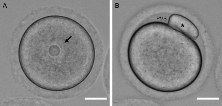
Figure 1.
Morphological differences between an oocyte and an egg. Transmitted light images showing key differences between a mammalian (mouse) (A) oocyte and (B) egg. The egg was obtained following in-vitro maturation. The nucleus or germinal vesicle is highlighted by the arrow and the polar body is highlighted by the asterisk. Note the increased perivitelline space (PVS) in the egg relative to the oocyte. Scale bar = 20 µm.

