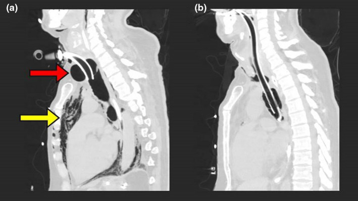
Figure 1.

(a) Sagittal computed tomography scan demonstrating tracheomegaly with tracheostomy cuff morphology and an anteroposterior diameter of 52.2 mm (red arrow), with associated severe pneumomediastinum (yellow arrow). (b) Sagittal computed tomography scan following oral tracheal re‐intubation below the defect, demonstrating the tracheal tube cuff in the lower trachea and a reduction in tracheal diameter.
