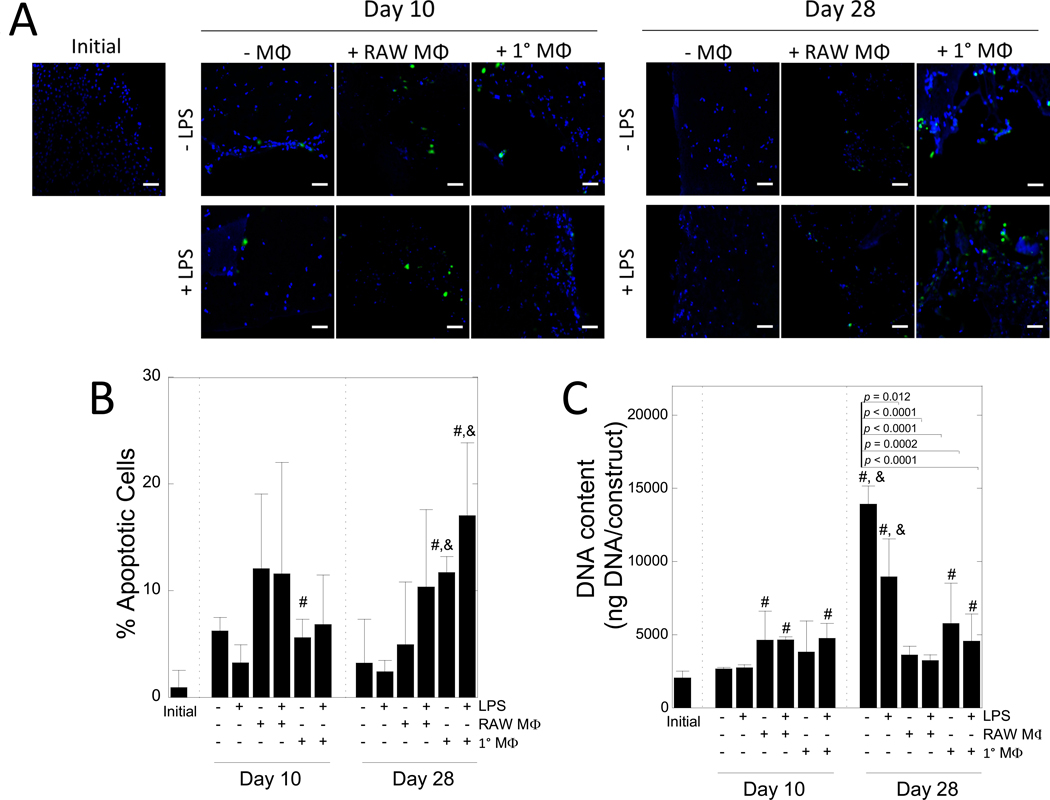
Figure 3 –
The effects of macrophages on MC3T3-E1 cell apoptosis. A) Representative confocal microscopy images of hydrogels immediately after encapsulation and after 10 and 28 days of culture. Cells were stained for apoptosis (green) and counterstained with DAPI for cell nuclei (blue). Scale bar = 50 μm. B) Semi-quantification of the percent of apoptotic cells, normalized to number of nuclei in hydrogels after 0, 10, or 28 days of culture. C) DNA content per construct over time. Data are shown as mean (n=3–4) with standard deviation as error bars. “#” denotes statistical significance (p < 0.05) over day 0, “&” denotes statistical significance (p < 0.05) over day 10.

