FIG 4.
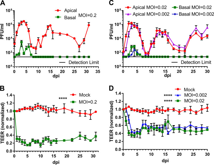
Virus release kinetics and transepithelial electrical resistance (TEER) measurement of HAE-ALI infected with SARS-CoV-2 at various viral loads (multiplicities of infection [MOIs]). (A and C) Virus release kinetics. HAE-ALIB4-20 cultures were infected with SARS-CoV-2 at an MOI of 0.2 (A) and 0.02 and 0.002 (C), respectively, from the apical side. At the indicated days postinfection (dpi), 100 μl of apical washes by incubation of 100 μl of D-PBS in the apical chamber and 100 μl of the basolateral media were taken for plaque assays. Plaque-forming units (PFU) were plotted to the dpi. Values represent means ± standard deviations. (B and D) TEER measurement. The TEER of mock- and SARS-CoV-2-infected HAE-ALI culture was measured using an epithelial volt-ohm meter (Millipore) at the indicated dpi. The TEER values were normalized to the TEER measured on the day of infection, which is set at 1.0. Values represent the means of relative TEER ± standard deviations. ****, P < 0.0001 by one-way Student’s t test.

