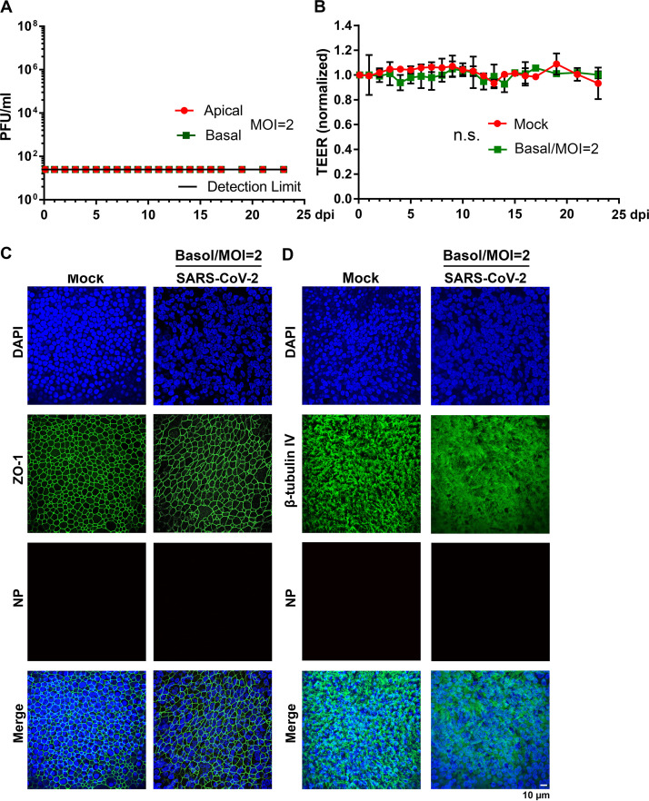
FIG 6.
SARS-CoV-2 does not infect HAE-ALI from the basolateral side. (A) Virus release kinetics. Both apical washes and basolateral media were collected from SARS-CoV-2-infected HAE-ALIB4-20 every day and quantified for virus titers using plaque assays. Plaque-forming units (PFU) were plotted to the dpi. Value represent means ± standard deviations. (B) TEER measurement. The TEER of infected HAE-ALIB4-20 cultures was measured using an epithelial volt-ohm meter (Millipore) at the indicated dpi, and were normalized to the TEER measured on the first day, which is set at 1.0. Values represent means of the relative TEER ± standard deviations. n.s., statistically not significant. (C and D) Immunofluorescence analysis. Mock- and SARS-CoV-2-infected HAE-ALIB4-20 cultures at 23 dpi were costained with anti-NP and anti-ZO-1 antibodies (C) or costained with anti-NP and anti-β-tubulin IV antibodies (D). Confocal images were taken at a magnification of ×40. Nuclei were stained with DAPI (blue). Basol, basolateral.

