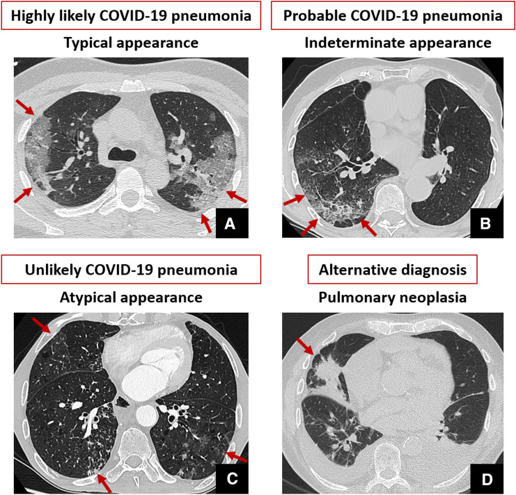
Fig. 1.
Exemplifying CT findings for each class of COVID-19 pneumonia probability based on the presence of typical, indeterminate and atypical finding, or eventual absence of signs of viral pneumonia and alternative CT diagnosis. A is reported unenhanced thin-section axial CT image showing typical peripheral ground-glass opacity with superimposed interlobular septal thickening and intralobular lines visibility (“crazy-paving” pattern), involving both lungs. Unilateral involvement, considered one of the indeterminate features, is reported in B. Atypical findings (C) include bronchiolar wall thickening and tree-in-bud opacities with centrilobular nodules (arrows in C). Finally, alternative diagnosis (D) was reported in the case of chest CT findings not typical for interstitial pneumonia and with clear pulmonary or extra-pulmonary findings explaining symptoms and laboratory test alteration (e.g., pulmonary neoplasia, red arrow in D, associated with bilateral pleural and pericardial effusion)

