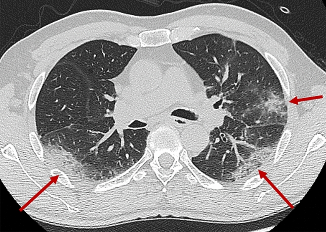
Fig. 3.

Chest CT typical COVID-19 pneumonia in a patient with initially negative swab. A 61-year-old man suffering from fever (39 °C), cough, and dyspnea from 7 days, presented to the emergency department of San Raffaele Hospital in Milan. Clinical evaluation and laboratory tests resulted highly suspicious for SARS-CoV2-related pneumonia. Nasopharyngeal swab and chest CT were immediately performed. CT showed peripheral opacity with crazy-paving pattern and consolidation (red arrows) mainly involving the upper left lobe and the lower lobes, mainly with posterior distribution. CT findings resulted highly suggestive for SARS-CoV2 pneumonia, but results from the first swab (available only 24 h later) resulted negative. In consideration of high clinical and CT suspicion, another swab was collected after 3 days, and it finally resulted positive
