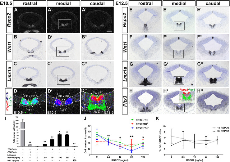
FIGURE 2.
RSPO2-mediated activation of WNT1/b-catenin signaling inhibits the differentiation of PITX3+ mdDA neurons in vitro. (A–H”) Representative coronal overviews (A–C”,E–H”) at different rostrocaudal levels of the midbrain (dorsal top) from CD-1 embryos at E10.5 (A–D’) and E12.5 (D”–H”), hybridized with riboprobes for Rspo2 (A–A”,E–E”), Wnt1 (B–B”,F–F”), Lmx1a (C–C”,G–G”), and Pitx3 (H–H”). (A–A”) dark-field images. (D–D”, inset in H’), pseudocolored overlays of consecutive medial midbrain sections (higher magnification dark- or bright-field views of boxed areas in A’–C’ and E’–H’, respectively), hybridized with probes for Rspo2 (red), Wnt1 (blue), and Lmx1a (green; D–D”) or Pitx3 (green; inset in H’), overlapping expression domains appear in magenta (red/blue), cyan (blue/green), or yellow (red/green). Broken white lines in (D–D”) outline the neuroepithelium and delimit lFP from mFP; white lines in (D”) delimit VZ/SVZ from IZ and MZ. (I) Fold change of luciferase (LUC) activity in HEK-293 cells (relative to only BSA-treated cells, set as 1) after transfection with TOPFlash or FOPFlash reporter and S33Y-b-catenin, or TOPFlash reporter and treatment with 10 μg BSA, increasing amounts of RSPO2 (2.5–250 ng/ml) or 100 ng/ml WNT1 protein [n = 3 independent experiments; statistical testing for significance between TOPFlash and FOPFlash + b-catenin (t = 5.8, df = 4, P = 0.0044, and unpaired t-test) or in relation to only BSA-treated cells]. (J) Quantification of PITX3+/TH– (green line), PITX3–/TH+ (red line), and PITX3+/TH+ (blue line) cells after treatment of VM primary cultures with BSA (10 μg) or increasing amounts of RSPO2 protein (2.5–100 ng/ml) for 7 days (10 DIV) [n = 5 independent experiments; statistical testing for significance only shown for double-positive cells in relation to BSA-treated cultures in two-way ANOVA for repeated measurements followed by Bonferroni’s posttests; F(4,36) = 6.28, P = 0.0024]. (K) Quantification of EdU+ per DAPI+ cells after treatment of VM primary cultures with BSA (10 μg) or increasing amounts of RSPO2 protein (2.5–100 ng/ml) for 1 day (4 DIV, gray line) or 3 days (6 DIV, black line) [n = 3 independent experiments; no significant change of EdU+/DAPI+ cells in relation to only BSA-treated cells in two-way ANOVA for repeated measurements followed by Bonferroni’s posttests; F(4,19) = 0.24]. *P < 0.05; **P < 0.005; and ns, not significant. Scale bar: 200 μm (A”).

