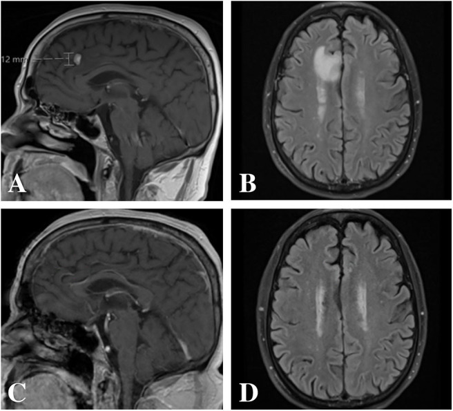
Figure 1.

Magnetic resonance imaging pre- and post-treatment. (A,B) Pre-treatment imaging demonstrated an avidly enhancing 11 × 7 × 12 mm lesion along the mesial surface of the right frontal lobe within the cingulate sulcus with surrounding vasogenic edema. (C,D) Post-treatment imaging with complete radiographic resolution of the lesion and associated vasogenic edema.
