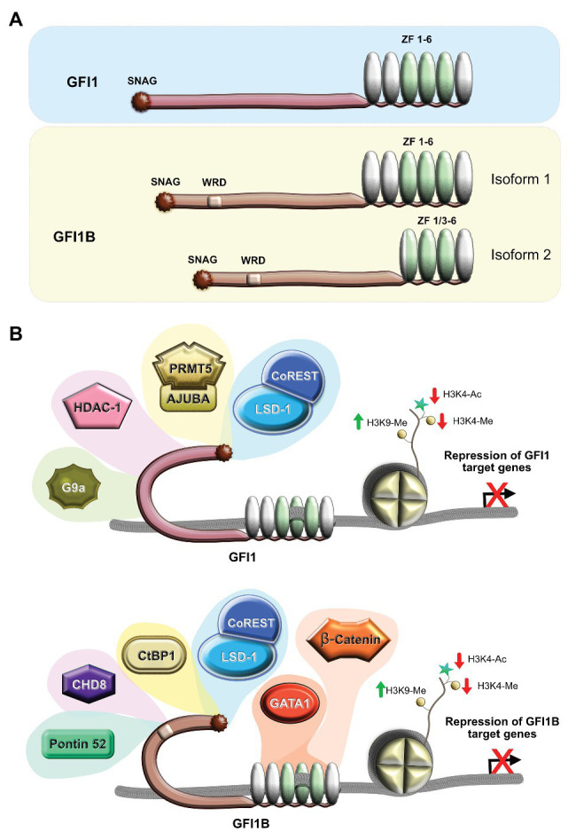
Figure 1.

Structure and function of human GFI1 and GFI1B. (A) Schematic depiction of the structure of the proteins, showing the SNAG suppressor domain, the less characterized intermediate domain involved in protein/protein interactions, and the six zinc finger domains (ZF) localized at the C-terminal end with those three involved in DNA binding shown in green and the three other domains that play a role in interaction with other proteins shown in silver. The two isoforms of Gfi1b are shown with the longer megakaryocyte-specific isoform 1 that has the six zinc fingers and the short erythroid-specific isoform 2 that lacks two zinc fingers due to the fusion of ZF1 and ZF3. (B) Schematic representation of the GFI1 (top) and GFI1B (bottom) complexes with different partners that promote gene silencing by removal of open chromatin signatures and induction of marks that correlate with closed chromatin. WRD, Wnt regulatory domain.
