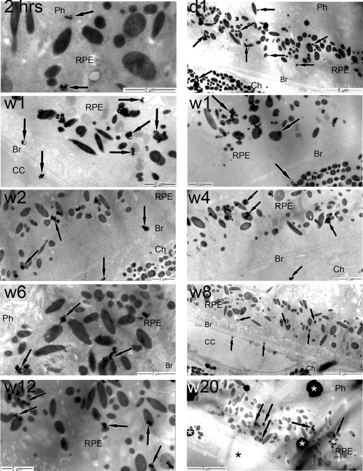
Figure 2.

Time course TEM autoradiographic distribution of 3H‐remofuscin (indicated by the presence of silver grains) in the posterior pole of the eye following a single intravitreal administration. The magnification calibration in micrometers is indicated in each electron micrograph. The black arrows show the silver grains, the asterisks indicate artefacts. Br, Bruch's membrane; CC, choriocapillaris; Ch, choroid; d, day; hrs, hours; Ph, photoreceptors; RPE, retinal pigment epithelium; w, week
