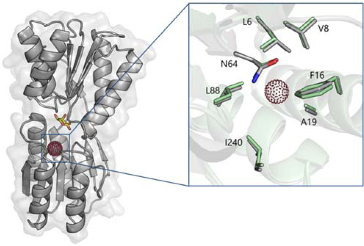
Figure 1.

Proposed Xe binding site in RBP(L19A). The protein model is based on the crystal structure of ribose-bound RBP in its closed conformation (PDB ID 2DRI). Xe (red dots) was modeled at the center of the cavity created by the L19A mutation. Bound ribose shown as yellow sticks. (Inset) Close-up view of the Xe binding site of RBP(L19A) in its closed (gray) and open (green; PDB ID 1URP) conformations.
