Figure 1.
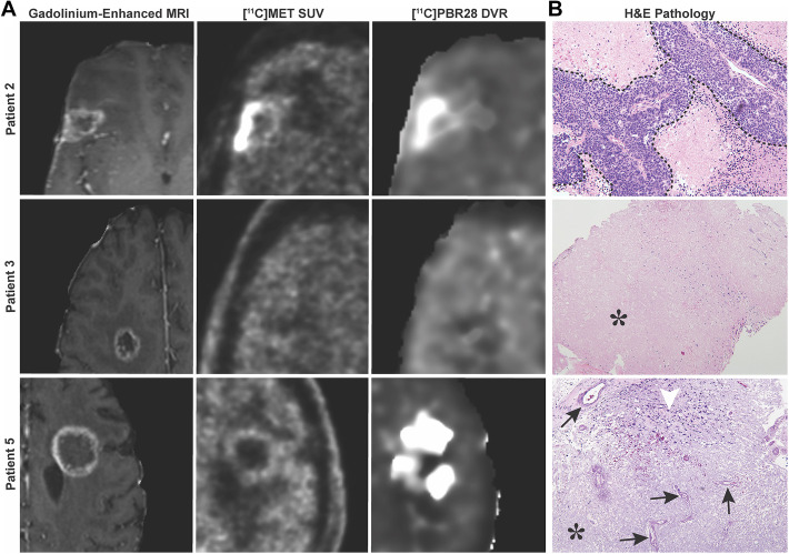
MRI and PET imaging for [11C]MET and [11C]PBR28. A, Representative images of suspicious lesions for TR or RN as seen on post-gadolinium MRI sequences. Corresponding lesions as seen on PET for [11C]MET and [11C]PBR28. B, Patient 2 had histologically confirmed TR, but had uptake of both [11C]MET and [11C]PBR28 radiotracers. Patient 3 had histologically confirmed RN but had absent uptake of both [11C]MET and [11C]PBR28 radiotracers. Patient 5 had histologically confirmed RN and demonstrated uptake of only [11C]PBR28. Tumor is outlined in a dashed line (top photo, taken at 20X). Characteristic features of RN include vessel hyalinization (arrows), increased immune infiltrate (arrowhead) (bottom photo, taken at 10X), and paucicelluar coagulative necrosis (*) (middle photo, taken at 4X, and bottom photo).

