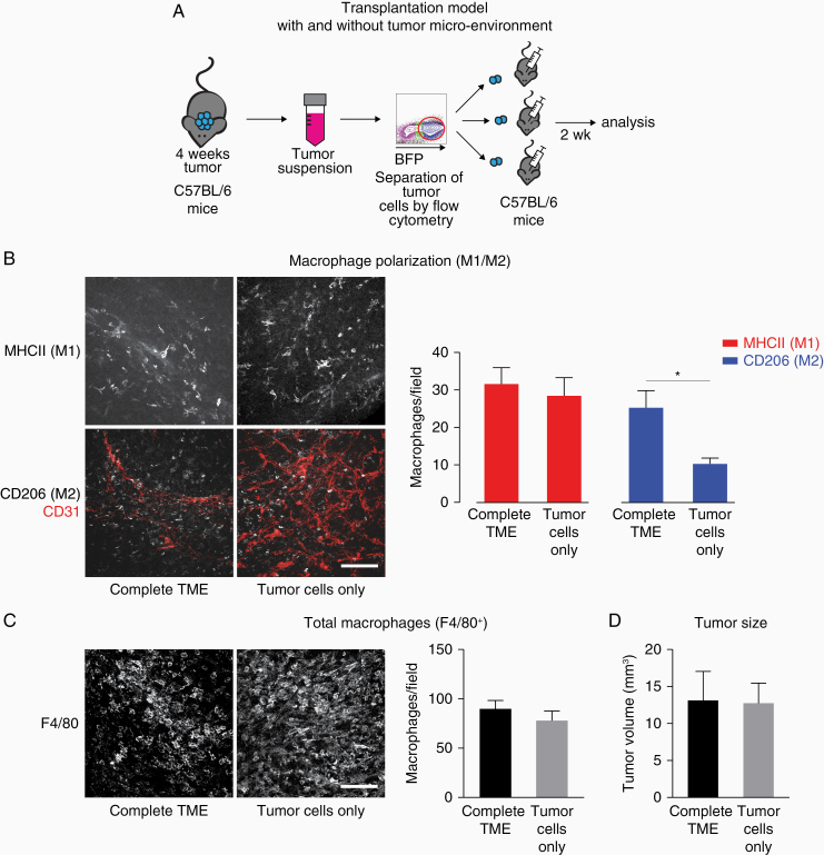
Figure 3.
The faster switch in macrophage polarization in the secondary tumors is mediated by the transplanted TME. (A) Schematic depicting the experimental approach. (B) Immunofluorescent staining of sections of 4 + 2 weeks tumor from complete TME and tumors from tumor cells only. Scale bar: 100 µm; 50 µm z-stack. Quantification of M1- and M2-positive macrophages. N = 10 (both groups); 5 fields of view/tumor. *P < .05. (C) Immunofluorescent staining and quantification of F4/80+ macrophages in 4 + 2 weeks tumor from complete TME and tumors from tumor cells only. Scale bar: 100 µm; 50 µm z-stack. N = 10 (complete TME) and 9 (tumor cells) tumors; 5 fields of view/tumor. (D) Tumor size of 4 + 2 weeks tumors from complete TME and tumors from tumor cells only. N = 10 (complete TME) and 8 (tumor cells) tumors. Data shown: mean + SEM (B–D).

