
Figure 4. Anti-CXCR3 treatment reduces TNF-α expression and elevates claudin-1 and AQP5 expression in the SMGs.
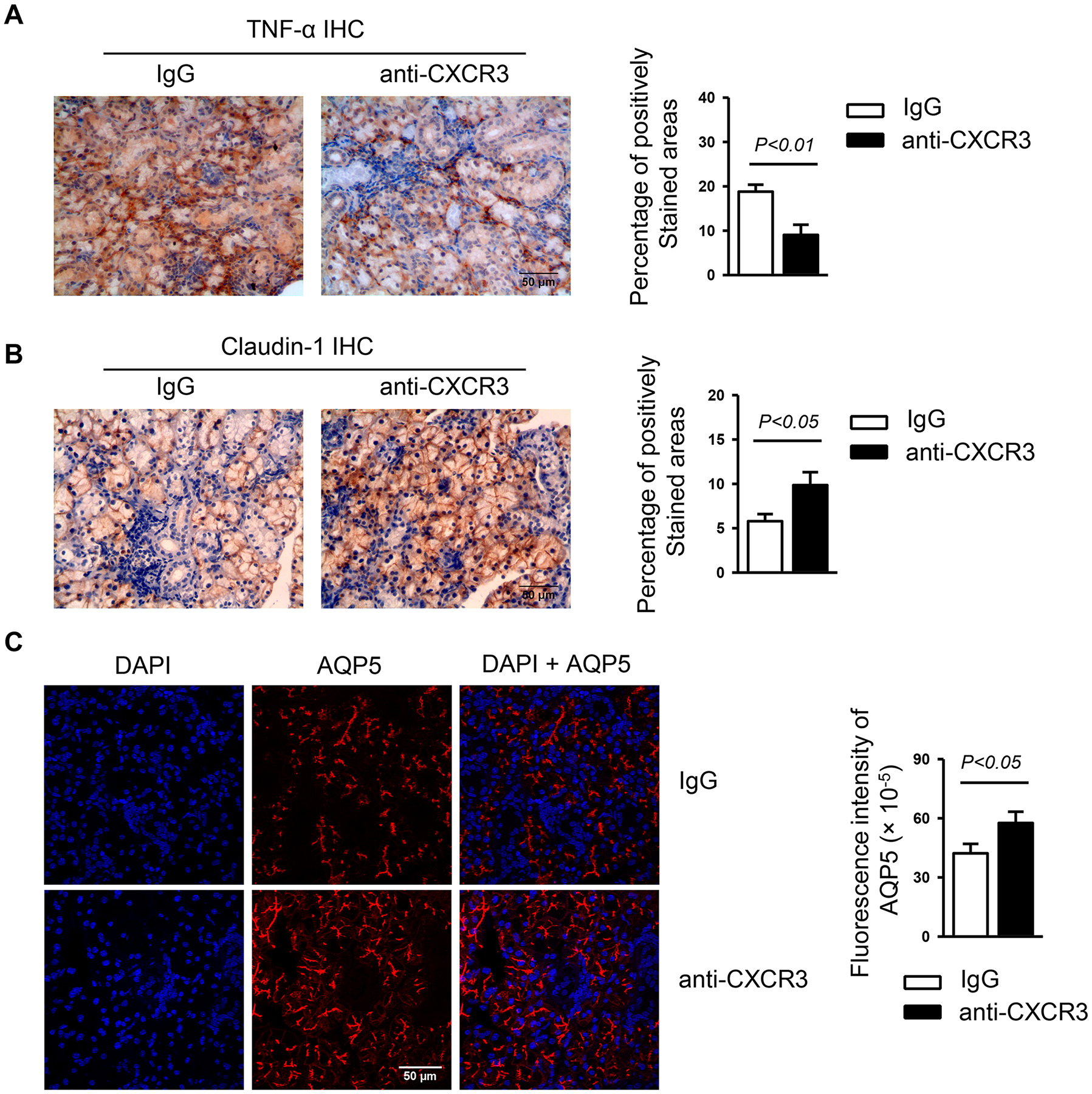
Anti-CXCR3 antibody or IgG was i.p.-administered to 4-week-old female NOD mice 3 times weekly for 6 weeks. (A) Immunohistochemical staining of TNF-α protein in SMG sections. Bar graph shows the percentage of positively stained areas in the sections. (B) Immunohistochemical staining of claudin-1 protein in SMG sections. Bar graph shows the percentage of positively stained areas in the sections. (C) Immunofluorescence staining of AQP5 protein in SMG sections. Bar graph shows the fluorescence intensity of AQP5 staining. Data are representative or the average of analyses of 12–15 mice for each group. All images were captured with ×400 original magnification.