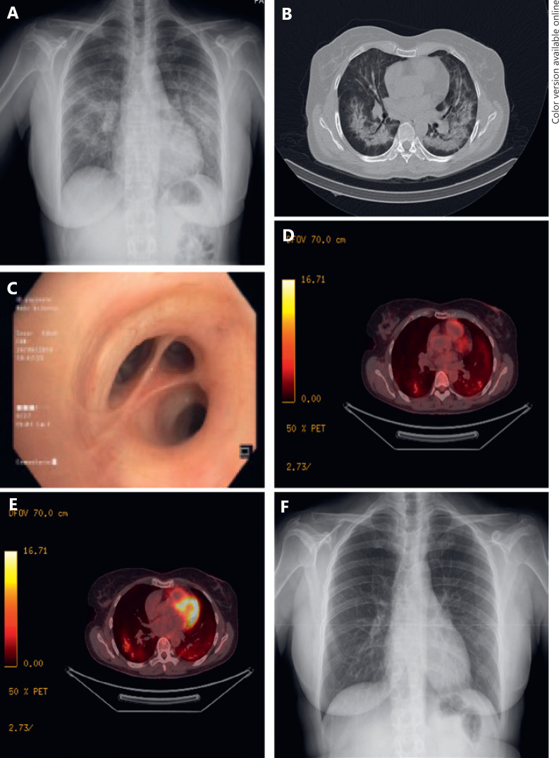
Fig. 1.
Radiological and pulmonological tests performed on our patient. A X-ray performed when the patient was admitted to the Emergency Department, showing a diffuse bilateral consolidation pattern. B CT performed during hospital admission, showing dense infiltrates in both lungs and areas with ground-glass attenuation. C Bronchoscopy showing no morphological alterations. D, E Positron-emission tomography-CT revealing a bilateral inflammatory pattern. F X-ray performed after 6 weeks with corticoid therapy showing complete resolution of the pulmonary infiltrations.

