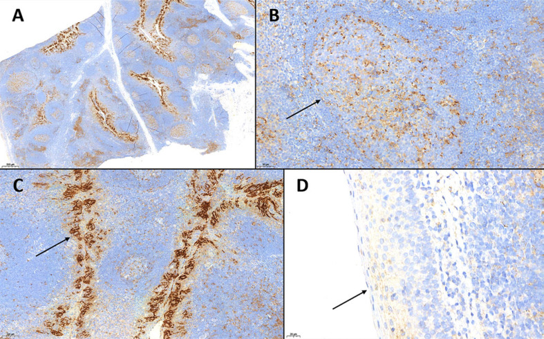
Fig. 1.
A PD-L1 IHC of a tonsil used as positive control. Note the tonsil parenchyma containing physiologically both PD-L1-positive immune cells (lymphocytes and monocytes), dispersed in the paracortical region (B) or in clusters within the germinal centers (B, arrow), and positive epithelial cells of the tonsil crypts (C). D The surface epithelium does not show any PD-L1 expression (each PD-L1 IHC, clone SP263., A ×20. B ×200. C ×100. D ×400.

