Fig. 1.
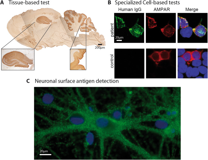
Example of comprehensive aurtoantibody testing available in scientiifc research settings. a Tissue-based test using sagittal rat brain sections optimized for detection of neuronal surface antibodies. Brown staining indicates specific binding of human IgG. Hippocampal and cerebellar regions are shown magnified. Shown is an example of the staining obtaining with GABA(A) receptor antibody containing patient CSF. b Cell-based assay with HEK293 cells expressing human autoantigens. This example shows cells transfected with AMPA receptor subunits stained with serum of a patient with anti-AMPA receptor encephalitis. Green staining indicates human IgG, red staining indicates a commercial antibody detecting transfected cells. Merged images are shown on the right side. Yellow indicates cells transfected and detected by human IgG thus indicating presence of autoantibodies targeting the transfected antigen. In addition to the shown example of cells fixed with paraformaldehyde, for some autoantibodies non-fixed live-cell based assays, which stain cells before addition of fixatives are more sensitive for the detection of some autoantibodies (e.g. MOG, not shown). c Primary, embryonal, rat hippocampal neurons are stained with serum from a patient with anti-AMPA receptor encephalitis. Cells are alive and non-permeabilized so that only surface antigens are detected by autoantibodies. Green indicates human IgG binding to individual synapses, blue is a DAPI counterstaining of nuclei to demonstrate presence of neurons

