Fig. 7.
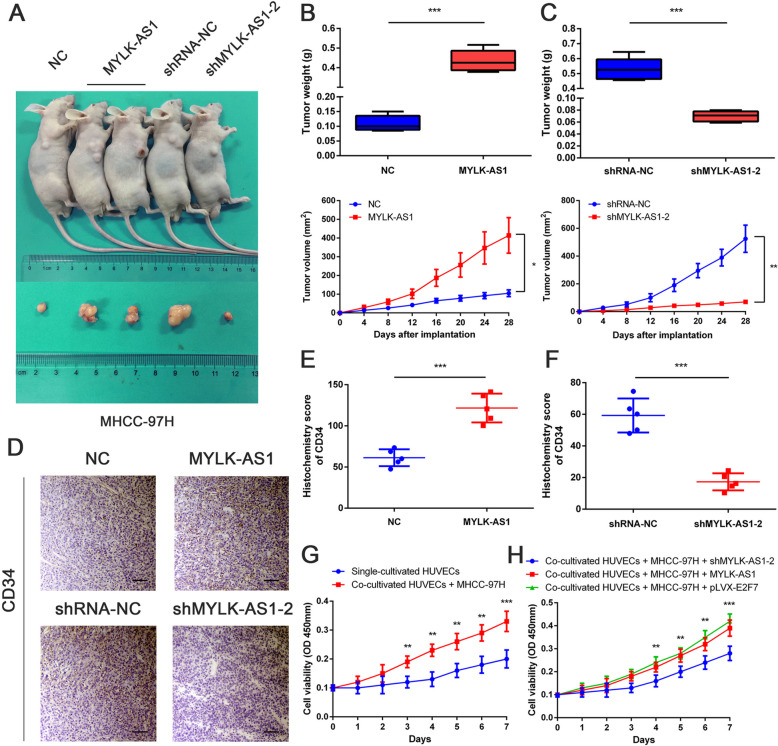
MYLK-AS1 regulates HCC cell proliferation and angiogenesis in vivo and in vitro. a Injection in the right armpit of MHCC-97H cells transfected with empty vector or MYLK-AS1 expression vector and shMYLK-AS1-NC or shMYLK-AS1–2 in the upper panel. Representative images of xenograft tumors are shown in the bottom panel. b-c Tumor weight and volume of the xenograft in MYLK-AS1 overexpression groups and control group or MYLK-AS1 knockdown group and control group. d Representative IHC staining results of CD34 in corresponding xenografts (scale bar = 50 μm). e-f Statistical analysis of the H-score of CD34 in the corresponding xenografts. Results are presented as mean ± SD from three independent experiments. g Cell proliferation of HUVECs cells cultured alone and co-cultured with MHCC-97H cells by CCK-8. h MYLK-AS1 knocked down or overexpressed or E2F7 overexpressed MHCC-97H cells co-cultured with HUVECs cells and consequent HUVEC proliferation by CCK-8. Results are expressed as mean ± SD. *P < 0.05, **P < 0.01, and ***P < 0.001

