Fig. 1.
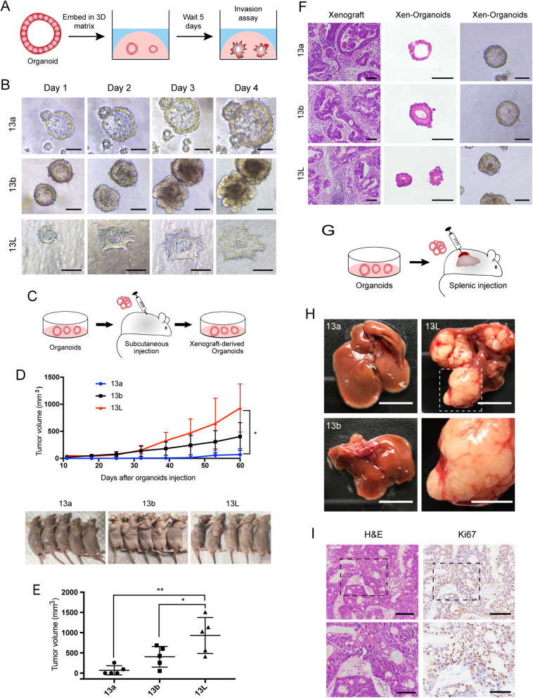
Patient-derived paired organoids provide invasion and transplantable models for human CRC progression. a Schematic of 3D invasion assay using paired organoids. b Representative micrographs of organoids in 3D invasion assay. Tumor organoids showed the smooth and protrusive leading fronts, respectively. The scale bar represents 100 μm. c Schematic of the subcutaneous organoid injection. d Organoids were injected subcutaneously into the flank region of nude mice for 60 days. Tumor volumes were monitored over time (top). n = 5 mice per group. Error bars indicate SEMs. *p = 0.0146 (one-way ANOVA). e Tumor volume of the mice at day 60 after inoculation. Each dot indicates individual mice. n = 5 mice per group. Error bars indicate SEMs. *p = 0.0433, **p = 0.002 (one-way ANOVA). f Representative bright-field images of organoids together with H&E staining of xenografts generated from organoids, and organoids derived from the xenografts. The scale bar represents 100 μm. g Schematic of the hepatic metastasis assay by splenic organoid injection. h Representative macroscopic photographs of the whole liver. n = 5 mice per group. Top, scale bar of the whole liver represents 1 cm. Bottom, high magnification of inset. Scale bar, 5 mm. i Representative histopathology and Ki67 staining of liver metastatic lesions generated from splenic injection of 13L organoids. Top, the scale bar represents 100 μm; bottom, the scale bar represents 50 μm

